Biological Oxidation-Reduction
Redox potential – free energy changes
In reactions involving oxidation and reduction, the free energy change is proportionate to the tendency of reactants to donate or accept electrons. Free energy change expressed as oxidation-reduction or redox potential. The redox potential of a system is usually compared with the potential of the hydrogen electrode (0.0 volts at pH 0.0). However, for biologic systems, the redox potential is normally expressed at pH 7.0, at which pH the electrode potential of the hydrogen electrode is -0.42 volts. Enzymes involved in oxidation and reduction are called oxidoreductases and are classified into four groups: oxidases, dehydrogenases, hydroperoxidases, and oxygenases. Oxidases use oxygen as a hydrogen acceptor. Oxidases catalyze the removal of hydrogen from a substrate using oxygen as a hydrogen acceptor and form water or hydrogen peroxide as a reaction product.
Some oxidases contain copper
Cytochrome oxidase is a hemoprotein widely distributed in many tissues, having the typical heme prosthetic group present in myoglobin, hemoglobin, and other cytochromes. It is the terminal component of the chain of respiratory carriers found in mitochondria and transfers electrons resulting from the oxidation of substrate molecules by dehydrogenases to their final acceptor, oxygen. The enzyme is poisoned by carbon monoxide, cyanide, and hydrogen sulfide. It has also been termed cytochrome a3. It is now known that cytochromes a and a3 are combined in a single protein, and the complex is known as cytochrome aa3. It contains two molecules of heme, each having one Fe atom that oscillates between Fe3+ and Fe2+ during oxidation and reduction. Furthermore, two atoms of Cu are present, each associated with a heme unit.
Other oxidases are Flavoproteins
Flavoprotein enzymes contain flavin mononucleotide (FMN) or flavin adenine dinucleotide (FAD) as prosthetic groups. FMN and FAD are formed in the body from the vitamin riboflavin. FMN and FAD are usually tightly – but not covalently – bound to their respective apoenzyme proteins. Metalloflavoproteins contain one or more metals as essential cofactors. Examples of flavoprotein enzymes include L-amino acid oxidase, an FMN-linked enzyme found in kidney with general specificity for the oxidative deamination of the naturally occurring L-amino acids.
Dehydrogenases cannot use oxygen as a hydrogen acceptor
There are a large number of enzymes in this class. They perform two main functions:
1. Transfer of hydrogen from one substrate to another in a coupled oxidationreduction reaction. These dehydrogenases are specific for their substrates but often utilize common coenzymes or hydrogen carriers, eg, NAD+ (Figure 9.1). Since the reactions are reversible, these properties enable reducing equivalents to be freely transferred within the cell. This type of reaction, which enables one substrate to be oxidized at the expense of another, is particularly useful in enabling oxidative processes to occur in the absence of oxygen, such as during the anaerobic phase of glycolysis.
2. As components in the respiratory chain of electron transport from substrate to oxygen.
Many dehydrogenases depend on Nicotinamide Coenzymes
These dehydrogenases use nicotinamide adenine dinucleotide (NAD+) or nicotinamide adenine dinucleotide phosphate (NADP+)—or both—and are formed in the body from the vitamin niacin. These coenzymes are reduced by the specific substrate of the dehydrogenase and reoxidized by a suitable electron acceptor. They may freely and reversibly dissociate from their respective apoenzymes. Generally, NAD-linked dehydrogenases catalyze oxidoreduction reactions in the oxidative pathways of metabolism, particularly in glycolysis, in the citric acid cycle, and in the respiratory chain of mitochondria. NADP-linked dehydrogenases are found characteristically in reductive syntheses, as in the extramitochondrial pathway of fatty acid synthesis and steroid synthesis— and also in the pentose phosphate pathway.
Other dehydrogenases depend on Riboflavin
The flavin groups associated with these dehydrogenases are similar to FMN and FAD occurring in oxidases. They are generally more tightly bound to their apoenzymes than are the nicotinamide coenzymes. Most of the riboflavin-linked dehydrogenases are concerned with electron transport in (or to) the respiratory chain. NADH dehydrogenase acts as a carrier of electrons between NADH and the components of higher redox potential. Other dehydrogenases such as succinate dehydrogenase, acyl-CoA dehydrogenase, and mitochondrial glycerol- 3-phosphate dehydrogenase transfer reducing equivalents directly from the substrate to the respiratory chain. Another role of the flavin-dependent dehydrogenases is in the dehydrogenation of reduced lipoate, an intermediate in the oxidative decarboxylation of pyruvate and a-ketoglutarate. The electron transferring flavoprotein is an intermediary carrier of electrons between acyl- CoA dehydrogenase and the respiratory chain.
Cytochromes may also be regarded as dehydrogenases
The cytochromes are iron-containing hemoproteins in which the iron atom oscillates between Fe3+ and Fe2+ during oxidation and reduction. Except for cytochrome oxidase, they are classified as dehydrogenases. In the respiratory chain, they are involved as carriers of electrons from flavoproteins on the one hand to cytochrome oxidase on the other. Several identifiable cytochromes occur in the respiratory chain, ie, cytochromes b, c1, c, a, and a3 (cytochrome oxidase). Cytochromes are also found in other locations, eg, the endoplasmic reticulum (cytochromes P450 and b5), and in plant cells, bacteria, and yeasts.
Hydroperoxidases use hydrogen peroxide or organic peroxide as substrate
Two type of enzymes found both in animals and plants fall into this category: peroxidases and catalase. Hydroperoxidases protect the body against harmful peroxides. Accumulation of peroxides can lead to generation of free radicals, which in turn can disrupt membranes and perhaps cause cancer and atherosclerosis.
Peroxidases reduce peroxides using various electron acceptors
Peroxidases are found in milk and in leukocytes, platelets, and other tissues involved in eicosanoid metabolism. The prosthetic group is protoheme. In the reaction catalyzed by peroxidase, hydrogen peroxide is reduced at the expense of several substances that will act as electron acceptors, such as ascorbate, quinones, and cytochrome c. The reaction catalyzed by peroxidase is complex, but the overall reaction is as follows: In erythrocytes and other tissues, the enzyme glutathione peroxidase, containing selenium as a prosthetic group, catalyzes the destruction of H2O2 and lipid hydroperoxides by reduced glutathione, protecting membrane lipids and hemoglobin against oxidation by peroxides.
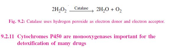
Catalase uses hydrogen peroxide as electron donor and electron acceptor
Catalase is a hemoprotein containing four heme groups. In addition to possessing peroxidase activity, it is able to use one molecule of H2O2 as a substrate electron donor and another molecule of H2O2 as an oxidant or electron acceptor (Figure 9.2). Under most conditions in vivo, the peroxidase activity of catalase seems to be favored. Catalase is found in blood, bone marrow, mucous membranes, kidney, and liver. Its function is assumed to be the destruction of hydrogen peroxide formed by the action of oxidases. Peroxisomes are found in many tissues, including liver. They are rich in oxidases and in catalase, Thus, the enzymes that produce H2O2 are grouped with the enzyme that destroys it. However, mitochondrial and microsomal electron transport systems as well as xanthine oxidase must be considered as additional sources of H2O2.
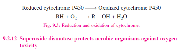
Cytochromes P450 are monooxygenases important for the detoxification of many drugs
Cytochromes P450 are an important superfamily of heme-containing monooxgenases, and more than 1000 such enzymes are known. Both NADH and NADPH donate reducing equivalents for the reduction of these cytochromes, which in turn are oxidized by substrates in a series of enzymatic reactions collectively known as the hydroxylase cycle (Figure 9.3). In liver microsomes, cytochromes P450 are found together with cytochrome b5 and have an important role in detoxification. Benzpyrene, aminopyrine, aniline, morphine, and benzphetamine are hydroxylated, increasing their solubility and aiding their excretion. Many drugs such as Phenobarbital have the ability to induce the formation of microsomal enzymes and of cytochromes P450. Mitochondrial cytochrome P450 systems are found in steroidogenic tissues such as adrenal cortex, testis, ovary, and placenta and are concerned with the biosynthesis of steroid hormones from cholesterol.
Reduced cytochrome P450 ?? Oxidized cytochrome P450 RH + O2 ?? R – OH + H2O
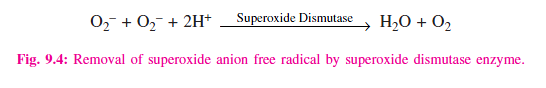
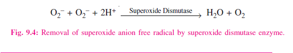
Superoxide dismutase protects aerobic organisms against oxygen toxicity
Transfer of a single electron to O2 generates the potentially damaging superoxide anion free radical (O2″-) (Figure 9.4), the destructive effects of which are amplified by its giving rise to free radical chain reactions. The ease with which superoxide can be formed from oxygen in tissues and the occurrence of superoxide dismutase, the enzyme responsible for its removal in all aerobic organisms (although not in obligate anaerobes) indicate that the potential toxicity of oxygen is due to its conversion to superoxide. Superoxide is formed when reduced flavins – present, for example, in xanthine oxidase – are reoxidized univalently by molecular oxygen.